Text and photos by Harold “Fritz” Moritz
| |
Amphiboles are important and widespread rock-forming minerals in the metamorphic rocks of
the highlands of Connecticut. Most of their specific characterization has been documented via
optical microscopy using thin-sections and published in bedrock geological quadrangle reports
since the 1950s. In less common instances, they can be found as large, distinct and collectible
crystals begging for detailed characterization. To date, most local amphiboles are identified as
tremolite (white to colorless), actinolite (green), hornblende (very dark green to black), or
anthophyllite (brown to green-brown in the Middletown Formation). The International
Mineralogical Association’s (IMA) many changes in nomenclature post-date most of the
literature about them in this state and so many more recognized species potentially exist based on
IMA’s subdivisions of earlier ones. To remedy this situation, during August 2016 and March
2017, I submitted samples of Connecticut amphiboles (or suspected ones) to Frank Craig for
TEM-EDS analyses. Frank is preparing an atlas of amphiboles and I am interested in
determining the various species found in Connecticut.
Amphiboles are extensive and complex group of minerals presently divided into a group/subgroup/root-name hierarchy and with either monoclinic (more common) or orthorhombic crystal symmetry. The latest IMA nomenclature report (Hawthorne et al., 2012) uses the general formula AB2C5T8O22W2 where:
A = ☐, Na, K, Ca, Pb2+
B = Li, Na, Mg, Fe2+, Mn2+, Ca
C = Li, Na, Mg, Fe2+, Mn2+, Zn, Co, Ni, Al, Fe3+, Cr3+, Mn3+, V3+, Ti, Zr
T = Si, Al, Ti
O = oxygen
W = O, OH, F, Cl
Individual members can often only be completely identified by a combination of chemical-analytical, X-ray diffraction and spectroscopic methods. However, TEM-EDS is a very powerful method that can by itself provide a wealth of chemical information to differentiate species, especially involving the A, B, C and T sites. In the list of elements above, the TEM-EDS method can cannot detect Li and H, so full characterization of the W site’s constituents is lacking and species cannot be split at that level. Li-containing amphiboles are rare and not expected in Connecticut. Nearly all amphiboles are monoclinic, so X-ray diffraction is rarely necessary so long as the samples are correctly visually identified as amphiboles, except in the case of differentiating the paramorphs anthophyllite (orthorhombic) from cummingtonite (monoclinic). But where that ambiguity exists, selected area electron dispersion (SAED) can be used.
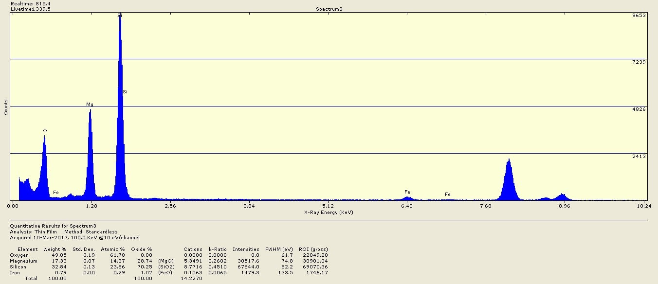
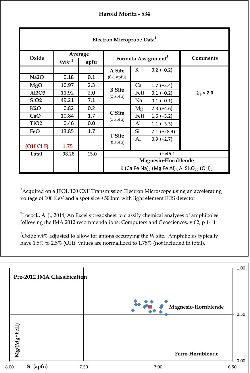
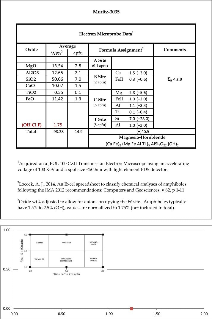
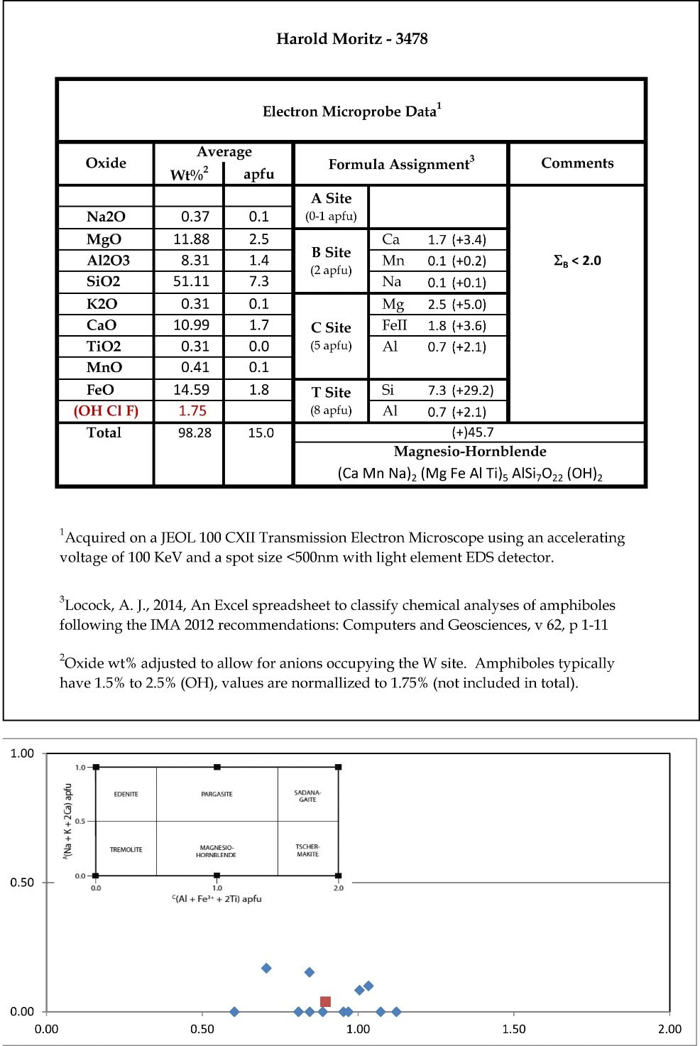
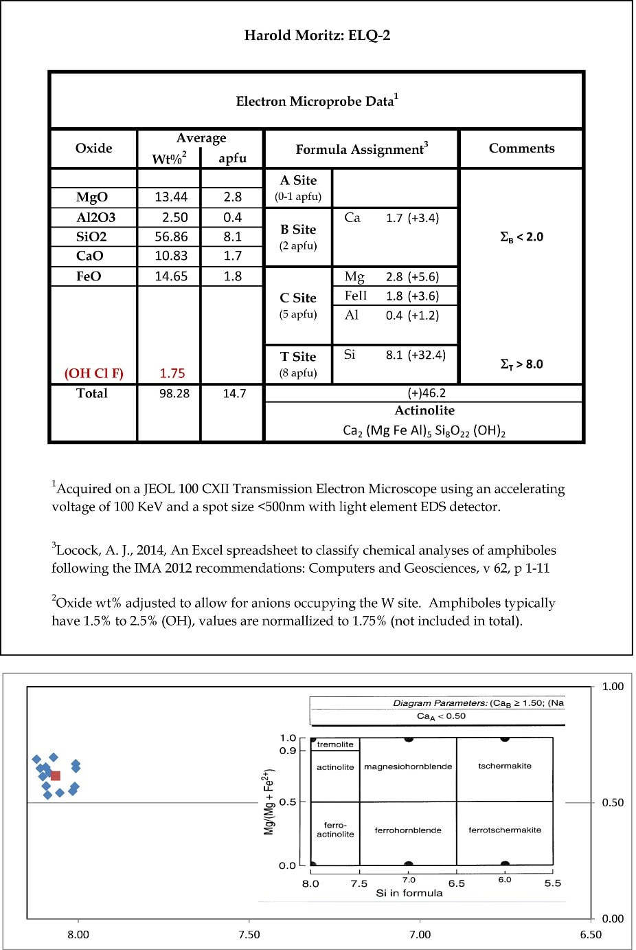
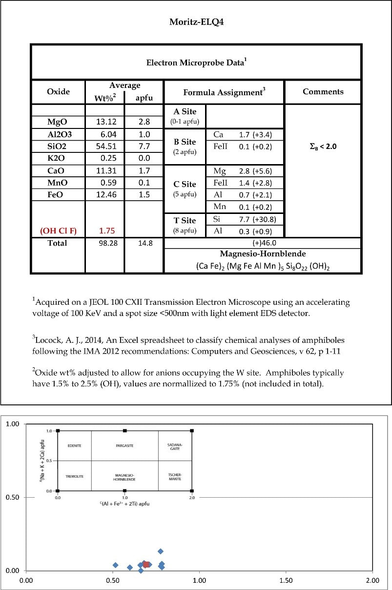
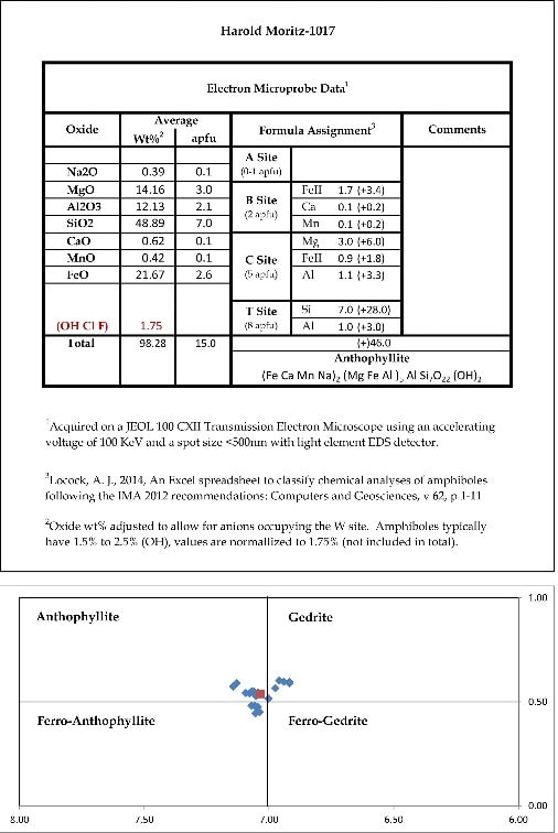
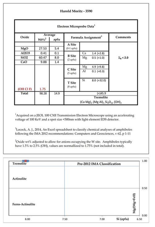
Below is a table of the samples, their suspected identification, the method used, and the results, followed by a discussion of each sample. The spectra were acquired by Frank Craig on a JEOL 100 CXII transmission electron microscope using an accelerating voltage of 100 KeV and a spot size <500 nm with light element EDS detector. Data reduction used Locock, A. J. (2014), An Excel spreadsheet to classify chemical analyses of amphiboles following the IMA 2012 recommendations. Computers and Geosciences: 62: 1-11. The oxide weight percent was adjusted to allow for anions occupying the W site. Amphiboles typically have 1.5% to 2.5% (OH), values are normalized to 1.75% (not included in total). Much of the resulting data have been included in the upcoming second edition of Frank Craig’s Asbestos Characterization, TEM Atlas of Regulated and Select Interference Minerals.
A = ☐, Na, K, Ca, Pb2+
B = Li, Na, Mg, Fe2+, Mn2+, Ca
C = Li, Na, Mg, Fe2+, Mn2+, Zn, Co, Ni, Al, Fe3+, Cr3+, Mn3+, V3+, Ti, Zr
T = Si, Al, Ti
O = oxygen
W = O, OH, F, Cl
Individual members can often only be completely identified by a combination of chemical-analytical, X-ray diffraction and spectroscopic methods. However, TEM-EDS is a very powerful method that can by itself provide a wealth of chemical information to differentiate species, especially involving the A, B, C and T sites. In the list of elements above, the TEM-EDS method can cannot detect Li and H, so full characterization of the W site’s constituents is lacking and species cannot be split at that level. Li-containing amphiboles are rare and not expected in Connecticut. Nearly all amphiboles are monoclinic, so X-ray diffraction is rarely necessary so long as the samples are correctly visually identified as amphiboles, except in the case of differentiating the paramorphs anthophyllite (orthorhombic) from cummingtonite (monoclinic). But where that ambiguity exists, selected area electron dispersion (SAED) can be used.
Below is a table of the samples, their suspected identification, the method used, and the results, followed by a discussion of each sample. The spectra were acquired by Frank Craig on a JEOL 100 CXII transmission electron microscope using an accelerating voltage of 100 KeV and a spot size <500 nm with light element EDS detector. Data reduction used Locock, A. J. (2014), An Excel spreadsheet to classify chemical analyses of amphiboles following the IMA 2012 recommendations. Computers and Geosciences: 62: 1-11. The oxide weight percent was adjusted to allow for anions occupying the W site. Amphiboles typically have 1.5% to 2.5% (OH), values are normalized to 1.75% (not included in total). Much of the resulting data have been included in the upcoming second edition of Frank Craig’s Asbestos Characterization, TEM Atlas of Regulated and Select Interference Minerals.
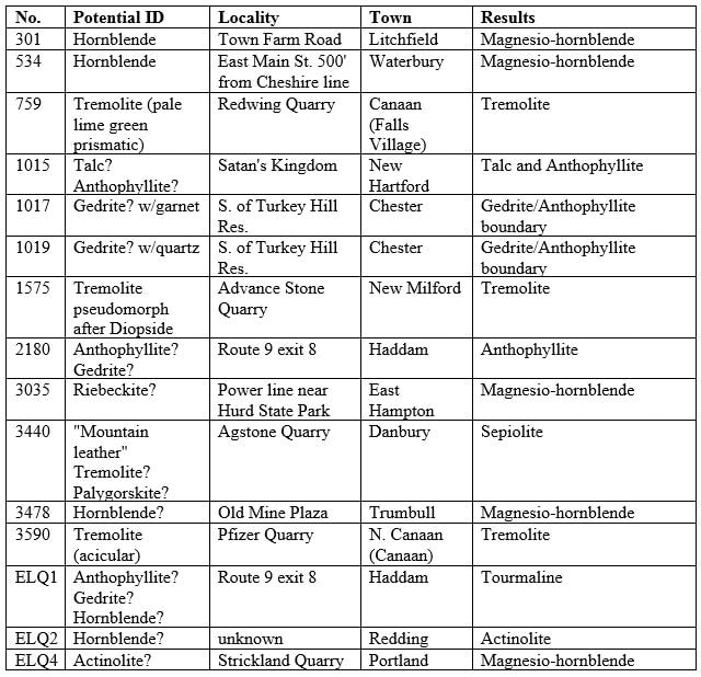
Samples
| Sample 301: A flabellate aggregate of “hornblende” crystals from the Town Farm Road area of northeast Litchfield, this fairly aesthetic specimen (12 x 10 x 5 cm) was in the John Schroder collection, I purchased it at the Connecticut Museum of Mining and Minerals in Kent. Similar crystallization occurs on Toll Gate Hill just south of US Route 202. Bedrock geology mapping shows interlayered amphibolites, amphibole gneiss, and schist underlying this area of the Cambro-Ordovician Rowe Formation. TEM-EDS analysis indicates it is magnesio-hornblende. | 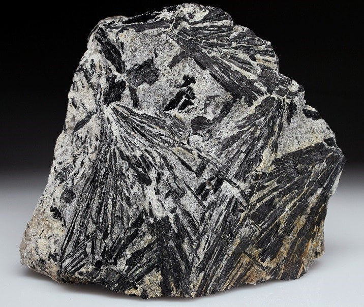 |

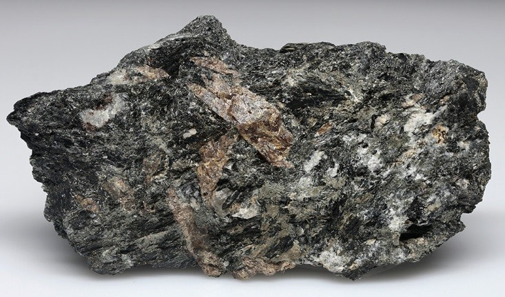
Sample 534: A dark greenish-black amphibole, probably a “hornblende” found at a construction site along East Main Street in Waterbury about 150 meters from the Cheshire line, in a calc-silicate rock associated with scapolite series and titanite. TEM-EDS results indicate it is magnesio-hornblende
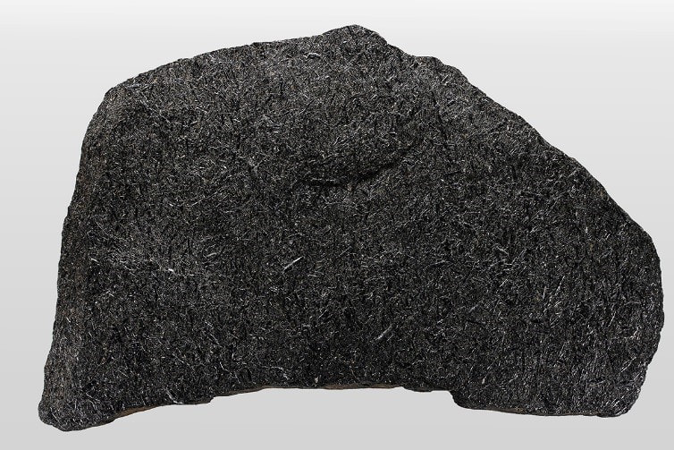
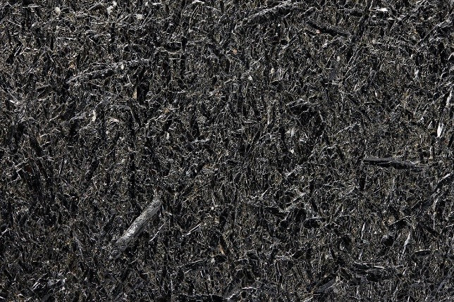
Sample 3035: A sample of amphibolite from the Middletown Formation collected by Richard Schooner from an outcrop along the power line right-of-way in East Hampton between Hurd and Seymour State Parks. He had labeled it as “riebeckite” likley because of its generally fine-grained nature (full view - 18.5 x 12 x 1.5 cm, close-up – FOV 6 cm). I wanted to confirm or refute this identification. The TEM-EDS results show that it is magnesio-hornblende.
 |  |
Sample 3478: A specimen of unaltered amphibolite hosting scheelite in the are of Old Mine Park in Trumbull, the TEM-EDS results indicates it is magnesio-hornblende.
| Sample ELQ-02: A sample of some dark greenish-black “hornblende” crystals from an unspecified locality in Redding, the specimen belongs to collector Eric L. Quinter, which he purchased some years ago. Photograph (FOV 5 cm) shows striated, flattened, elongated prisms, with golden chalcopyrite in quartz and calcite matrix (also with scapolite not visible). Apparently from a calc-silicate assemblage. In keeping with this paragenesis, the TEM-EDS results indicate it is actually actinolite. | 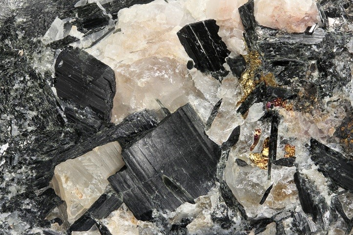 |
| Sample ELQ-04: A sample of acicular, dark greenish-black “actinolite” crystals with orange grossular and traces of massive red rutile. from a calc-silicate assemblage in the Collins Hill Formation adjacent to the Strickland pegmatite. The specimen belongs to collector Eric L. Quinter, which he purchased some years ago. This is identical to unanalyzed material described by Richard Schooner, who gave it the | 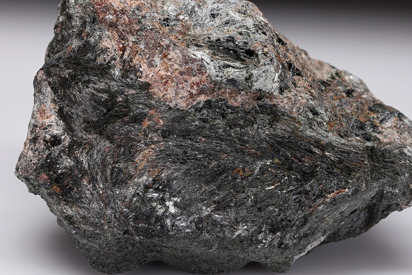 |
original identification. However, the TEM-EDS results indicate it is actually our friend magnesio-hornblende, although the subsamples do trend toward the actinolite field.
 | Sample 2180: In the 2000s, I collected a large amount of this aesthetic material (photographed one is 22 x 14 x 7 cm) from the DOT rock dump near the state Route 9 exit 8, Haddam before the state sold it off and residences were built on it. I was never really sure if it was gedrite or the reportedly more common anthophyllite because the geological report from 1979 does not include photographs |
of hand samples with mineral identifications. It does say that both are present as large crystals in the Middletown Formation, which is well exposed in Haddam and Chester. Anthophyllite is typically described as brownish, though the material I always see in the field is greenish, while gedrite is described as black. The TEM-EDS results indicate it is anthophyllite.
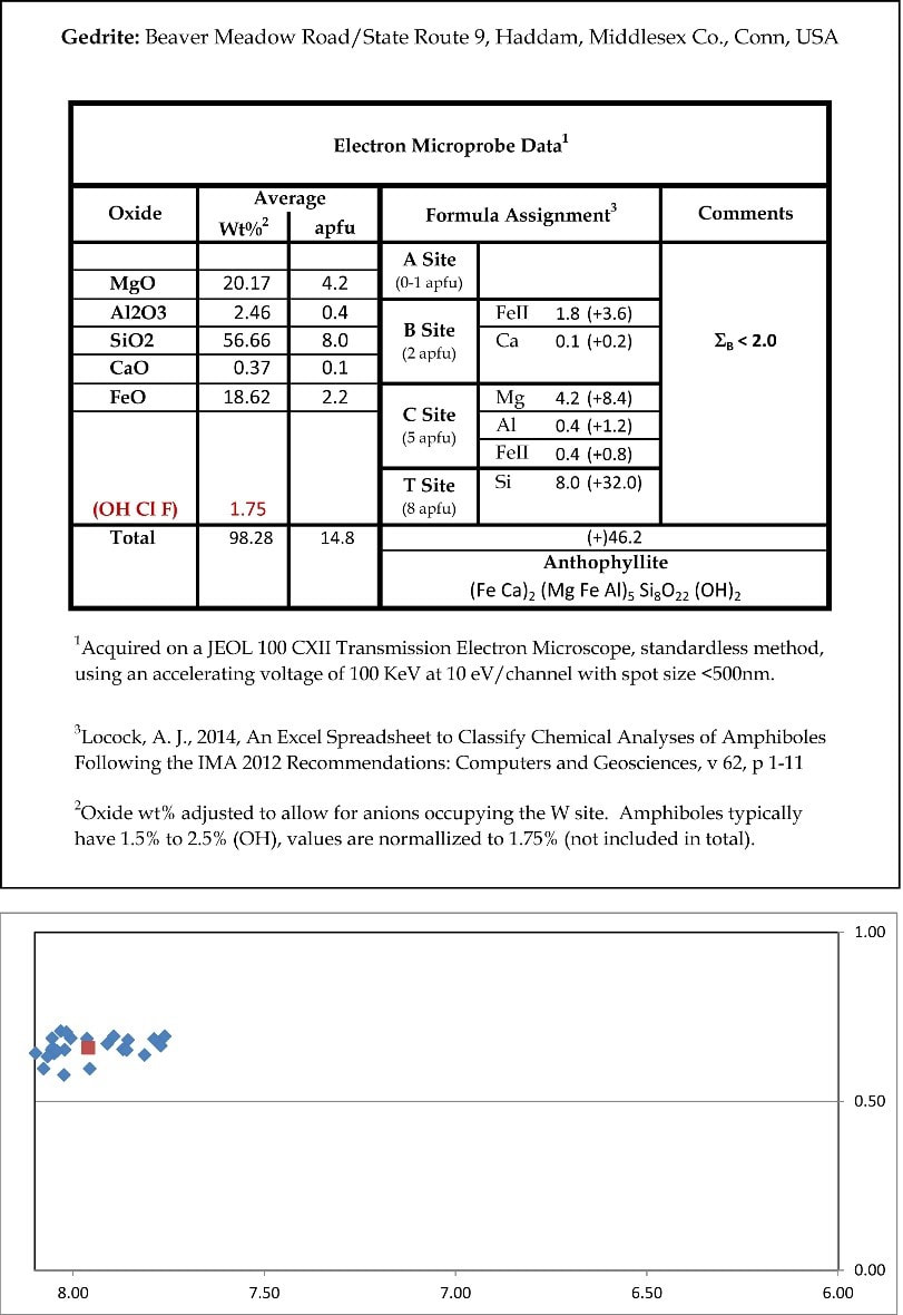
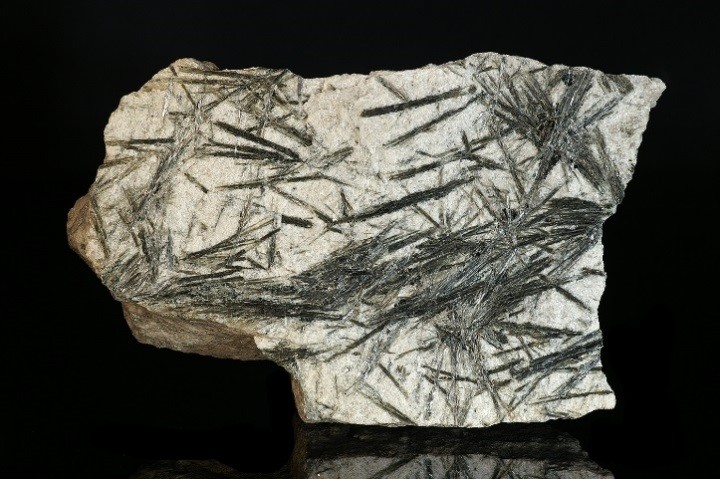
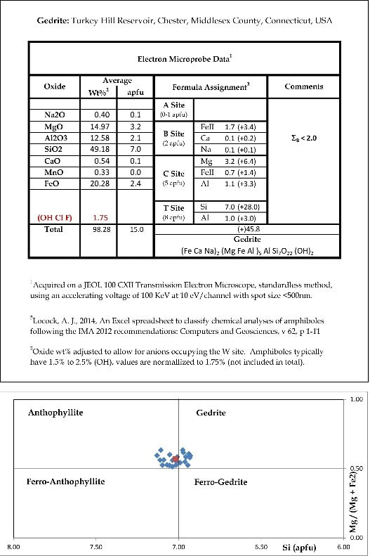
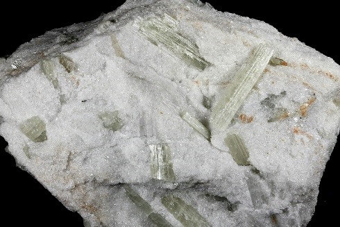
Samples 1017 and 1019: Searching for the gedrite described in the Lundgren’s 1979 Haddam Quadrangle bedrock geological report, in 2016 I went to an area “south of Turkey Hill Reservoir” where it states the area is “strewn with large blocks” of the lustrous, black “garnet/gedrite rock” – described as very coarse grained, with maximum crystals 5 to 10 cm. The area is strewn with broken down small outcrops and loose blocks of several kinds but eventually a dark rock with large reddish almandine (based on other EDS analyses) sticking through the moss and lichen-covered surface was located and sampled (full view photo - 8 x 7.5 x 3 cm). Right near the block was a small quartz rich boulder with similar large amphibole crystals, which was also sampled (close-up photo – FOV 13 cm).
 | 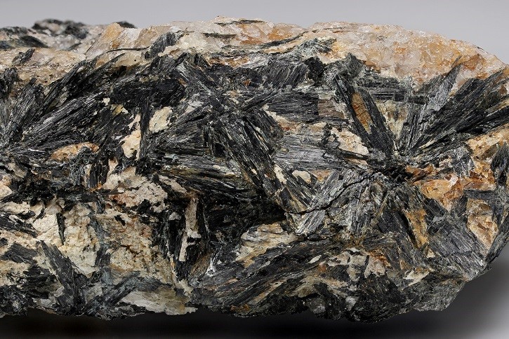 |
As indicated by the results from both samples, the average (red dot) of multiple subsamples of each is just barely in the anthophyllite compositional range, potentially rendering Lundgren’s 1979 identifications obsolete. The subsample data of sample 1017 trends into the adjacent gedrite and ferro-anthophyllite ranges, while 1019 trends into the gedrite range. Both just skirt the ferro-gedrite range. These crystals are essentially 4-way fence-sitters! Whether any of Lundgren’s “gedrite” crystals from the area are confirmed awaits further collecting and analyses.
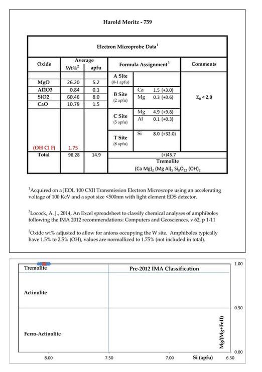
Samples 759, 1575 and 3590 (left to right, top to bottom): These are 3 sample of various habits of “tremolite” that are typical of the marble belt of western Connecticut, underlain by the Cambro-Ordovician Stockbridge/Inwood Marble. Based on their white to very pale lime green color, and historical information, there was little doubt that they are tremolite, one of the few amphiboles that can be reliably sight-identified. The goal was to confirm their identification with the same level of data quality as the other amphibole samples and to see if the composition varied along with the habit.
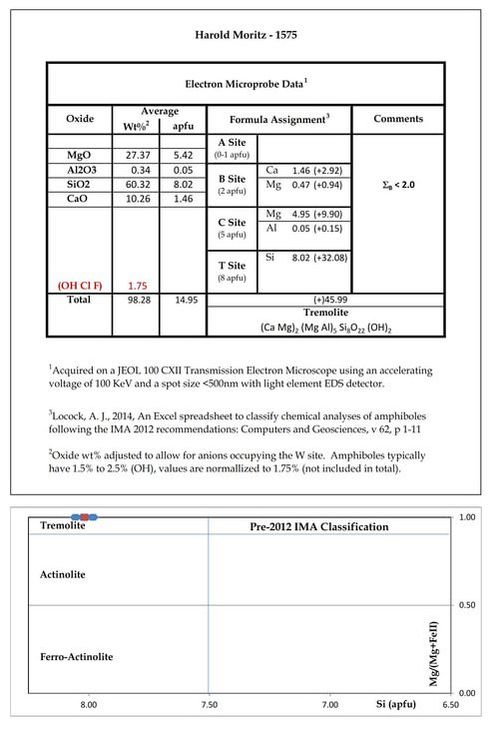
 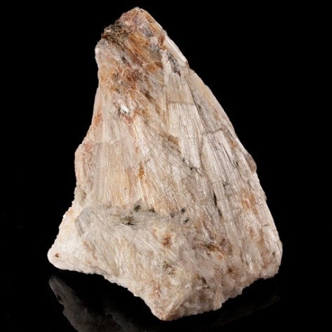 | 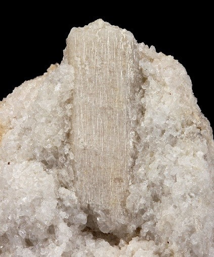 Sample 759 represents a typical pale lime green prismatic crystal from Redwing Quarry, Falls Village, Canaan. Sample 1575 is a tremolite after diopside “canaanite” pseudomorph from Advance Stone Quarry, New Milford, which are common all along the belt. The analyzed crystal is similar to |
the 26 mm crystal in the photograph.
Sample 3590 is from a typical splintery radiating crystal aggregate (8.5 x 5.5 x 5.5 cm) from Pfizer Quarry, Canaan village, North Canaan. This is also a very common tremolite habit in the marble belt, most of it occurs this way.
As expected, the TEM-EDS results confirmed the tremolite identification for all 3 samples.
| Sample 1015: These masses of very soft, fibrous, radiating, stellate material (FOV 33 mm) from a small quarry in New Hartford can be found in many collections. While preparing the text for the mindat.org page on it that I updated, I found that it has been called many minerals on labels and in various publications over the decades without any analytical data: | 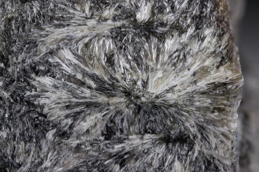 |
Actinolite: {Ca2}{Mg4.5-2.5Fe0.5-2.5}(Si8O22)(OH)2
Pyrophyllite: Al2Si4O10(OH)2
Talc: Mg3Si4O10(OH)2
Anthophyllite: {Mg2}{Mg5}(Si8O22)(OH)2
This ambiguity needed to be clarified. It seemed to me the whole package of visual and physical properties are most like the pyrophyllite deposits in North Carolina and Graves Mountain, Georgia.
Pyrophyllite: Al2Si4O10(OH)2
Talc: Mg3Si4O10(OH)2
Anthophyllite: {Mg2}{Mg5}(Si8O22)(OH)2
This ambiguity needed to be clarified. It seemed to me the whole package of visual and physical properties are most like the pyrophyllite deposits in North Carolina and Graves Mountain, Georgia.
Initially an SEM-EDS analysis was conducted courtesy of Micromounters New England with some ambiguous results. It did show, surprisingly, that it is definitely not pyrophyllite, there is no aluminum, and there is a lot of Mg present. It is some kind of Mg-Fe silicate, with the Smart Quant report indicating it is magnesium-dominant with a Mg/(Mg+Fe) ratio of 0.77. Based on the initial analytical results, it is possibly ferroan anthophyllite (Mg>Fe) or ferroan cummingtonite (Mg>Fe); or one of the similar but rarer non-aluminous amphiboles. However, given its softness, it could be talc. Clearly the limitations of SEM-EDS were reached on this mineral and that another type of analysis was necessary.
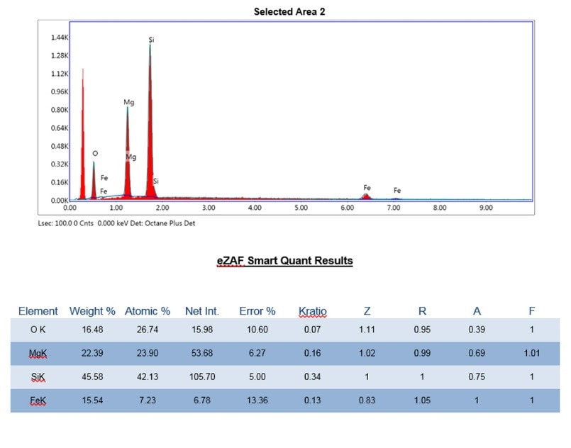
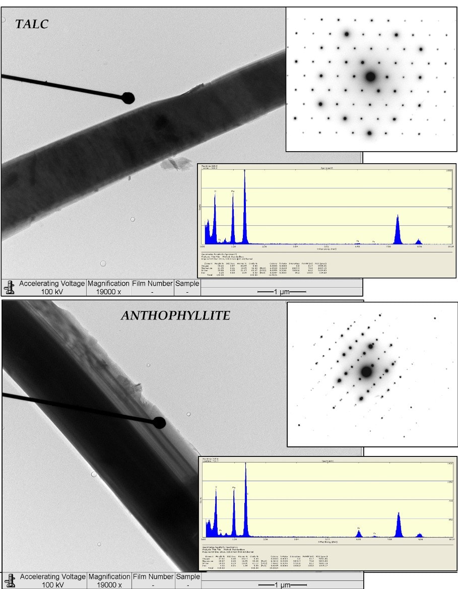
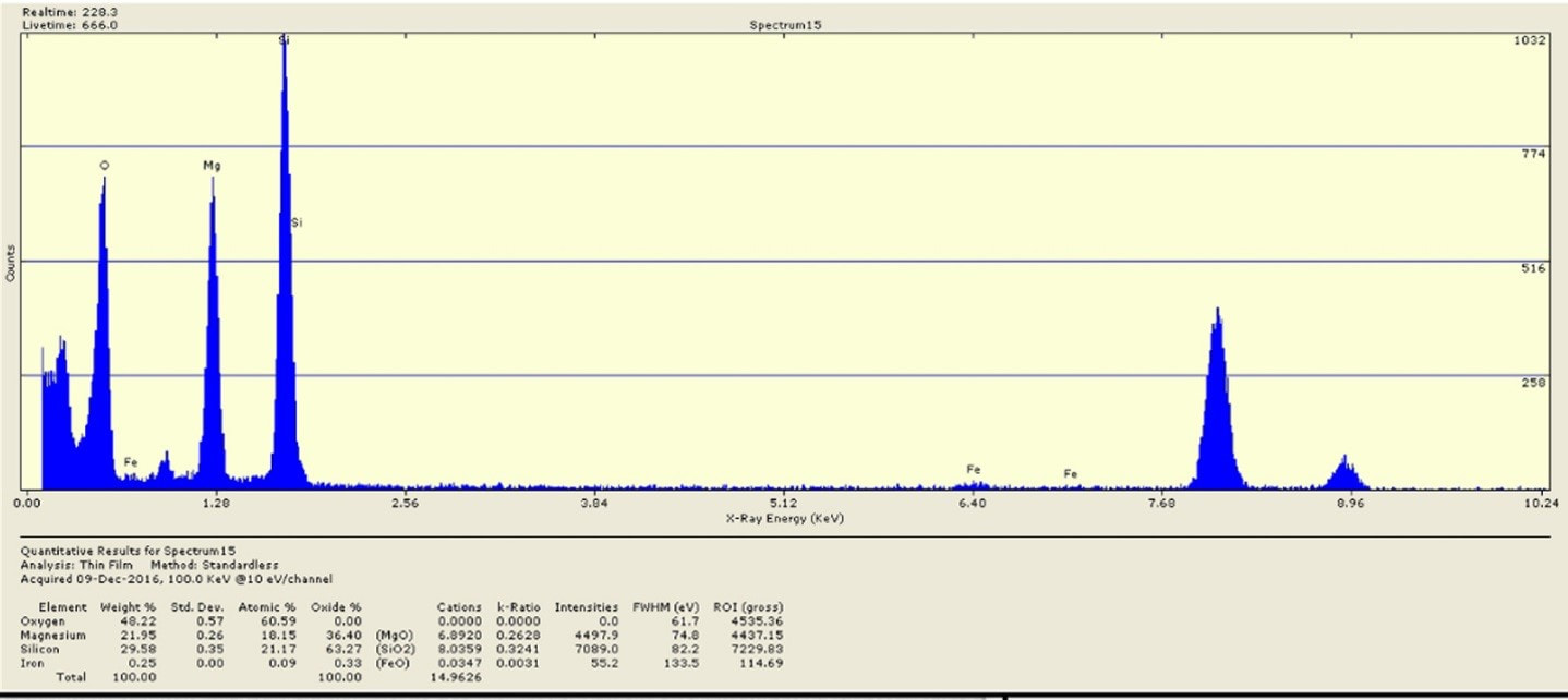
Therefore, optical microscopy, selected area electron diffraction (SAED) zone patterns and TEM-EDS analyses were conducted in 2016. Collectively, they show the material is essentially fibrous talc containing/contaminated with fibrous anthophyllite with a little Fe impurity. Note the similarity of the EDS spectra, but the SAED zone patterns show the difference in crystal structure. No wonder this was a tough nut to crack! Those results are shown below:
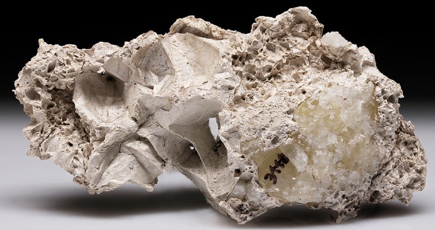
Sample 3440: A nice piece of “mountain leather” (11 x 5.5 x 2 cm) that formed sandwiched between 2 layers of calcite “dogtooth” habit crystals (note abundant “toothmarks”), found at the Agstone Quarry, Danbury and labeled “palygorskite” by Ronald Januzzi. “Mountain leather” is a field term for uncharacterized mats or masses of finely fibrous mineralization that could be tremolite, but could also be palygorskite, (Mg,Al)2Si4O10(OH)·4H2O or more likely sepiolite, Mg4Si6O15(OH)2 · 6H2O. The latter 2 minerals are not amphiboles, but have similar chemistry aside from the Al in palygorskite. The TEM-EDS results did not detect Al and are a match for sepiolite.