by Harold "Fritz" Mortiz

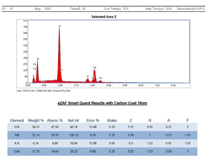
Apophyllite Specimen 816, O&G Quarry No. 2, Southbury/Woodbury
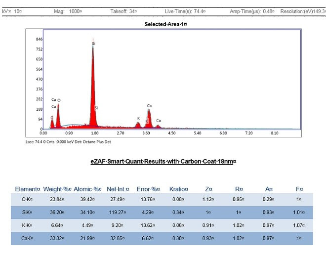
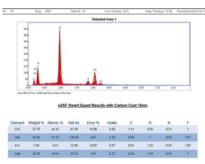
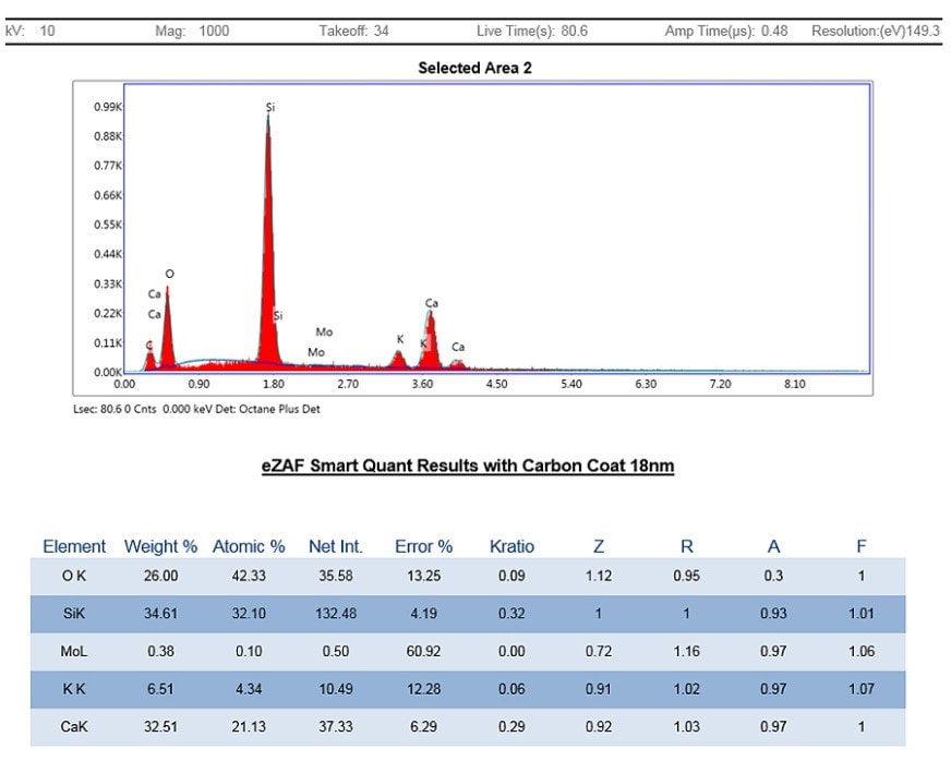
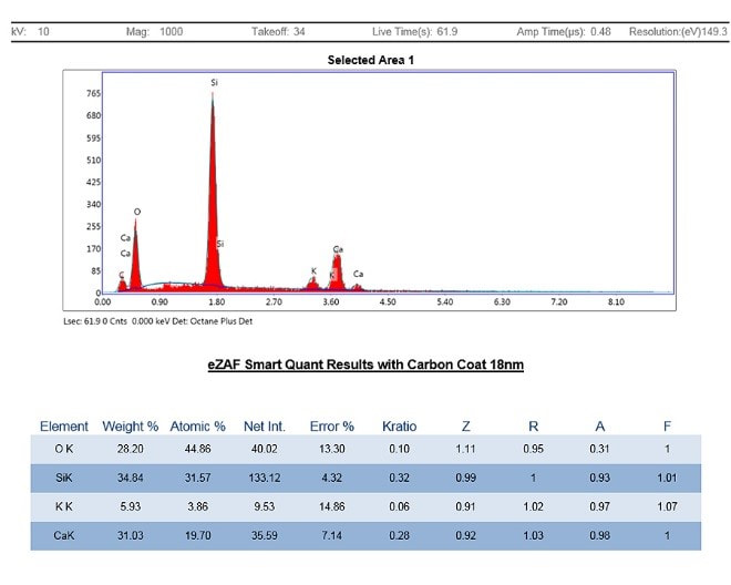
Recently I submitted samples of apophyllite from several places around Connecticut for analyses. Micromounters New England will perform a limited number of scanning electron microscope – energy dispersive spectroscopy (SEM-EDS) analyses for members such as me. SEM-EDS provides elemental chemistry on extremely small samples except for the very light elements H, He, B, Be and Li.
The goals of this project were to better determine the particular species of apophyllite on the specific samples and from that information hopefully extend the species identifications to unanalyzed specimens from the same localities, and perhaps throughout the state. Consequently, at least one sample was selected from most known localities with 3 samples submitted from the very prolific O&G locality.
The goals of this project were to better determine the particular species of apophyllite on the specific samples and from that information hopefully extend the species identifications to unanalyzed specimens from the same localities, and perhaps throughout the state. Consequently, at least one sample was selected from most known localities with 3 samples submitted from the very prolific O&G locality.
Originally described as one mineral in 1806, since 1978 the apophyllite group has been divided into 3 similar-looking species - fluorapophyllite-(K), fluorapophyllite-(Na), and hydroxyapophyllite-(K), with the general formula (K, Na)Ca4(Si8O20)(F,OH) · 8H2O. The particular species depends on the relative proportion of K and Na and/or F and OH. In general, according to mindat.org, fluorapophyllite-(K) is far more common than hydroxyapophyllite-(K) and fluorapophyllite-(Na) is very rare. However, some of the disparity may be due to a lack of analyses. Unfortunately, because of the limitations of SEM-EDS, the analyses would not be able to detect H (and detection of F is difficult), and therefore the ratio of F to OH could not be quantified. But at a minimum the dominance of K or Na would easily be found.
Below is a table of the samples (all from my collection) followed by a discussion of the results.
Below is a table of the samples (all from my collection) followed by a discussion of the results.
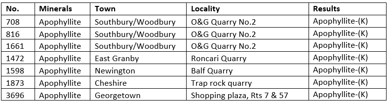
Apophyllite crystals are common in Connecticut only at a few places in the basalt flows (trap rock) where there is significant mineralization in gas vesicles and fractures. These are primarily the two O&G quarries in Woodbury (No. 1) and on the Woodbury/Southbury line (No. 2) and the Roncari Quarry in East Granby. It is also less commonly found at the quarry at Reed’s Gap on the Durham/ Wallingford line (a sample I submitted from there turned out to be the wrong mineral, so it is excluded from this discussion), rarely at the Balf Quarry, Newington and at Hamden’s old Pine Rock Quarry and the old trap rock quarry in Cheshire. In this geo-environment it typically occurs with pumpellyite, calcite, quartz, babingtonite and zeolite group minerals (stilbite, heulandite, analcime, natrolite, chabazite). Myriad attractive specimens have been saved from these localities.
Apophyllite can also rarely occur in fractures in metamorphic rocks, with one decent locality represented by the rock cut in Georgetown, where it occurs with spectacular mesolite crystals plus albite, heulandite and stilbite.
All of the SEM-EDS results shown below found the fluor/hydroxyapophyllite-(K) series, with no Na detected in any of them. Consequently, I think it is safe to conclude that any apophyllite found elsewhere in the state should belong to this series, unless it is identified at a new locality that has a very different mineral-forming geo-environment than the ones above. In that case, a full analysis should be done.
Specimen Analyses and Descriptions
| Specimen 708 Pinkish-white apophyllite-(K) with drusy, tabular datolite. FOV 5 cm tall. H. Moritz photo. | 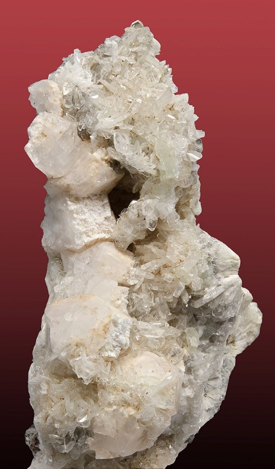 |
Specimen 816
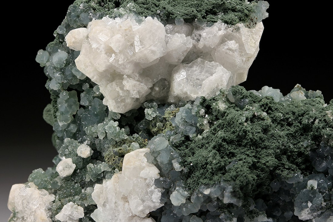
White apophyllite-(K) with greenish pumpellyite and blue-green prehnite. FOV 6 cm. H. Moritz photo.

Specimen 1472
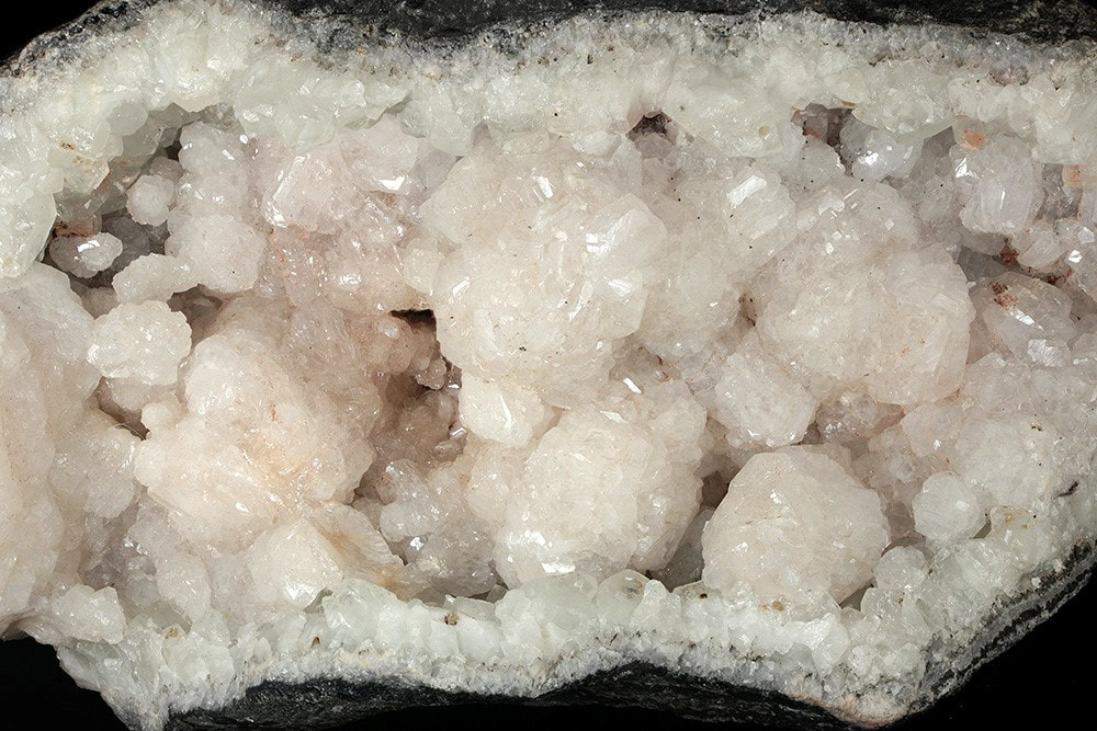 | 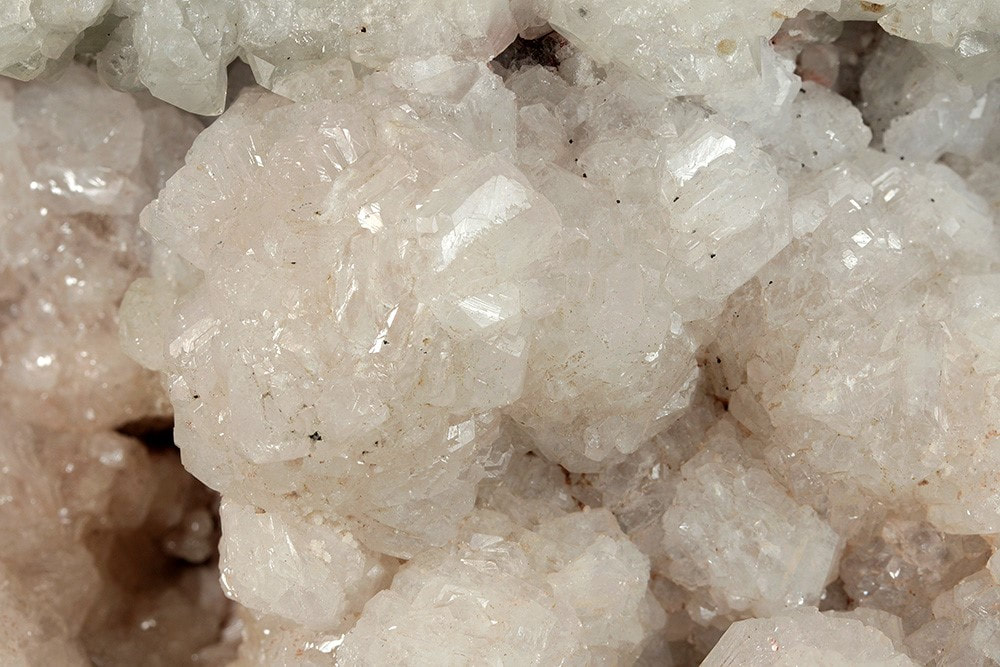 |
Spherical aggregates of tabular apophyllite-(K) crystals. FOVs – 14 cm tall (left), 7 cm tall (right). H. Moritz photo.
Specimen 1598
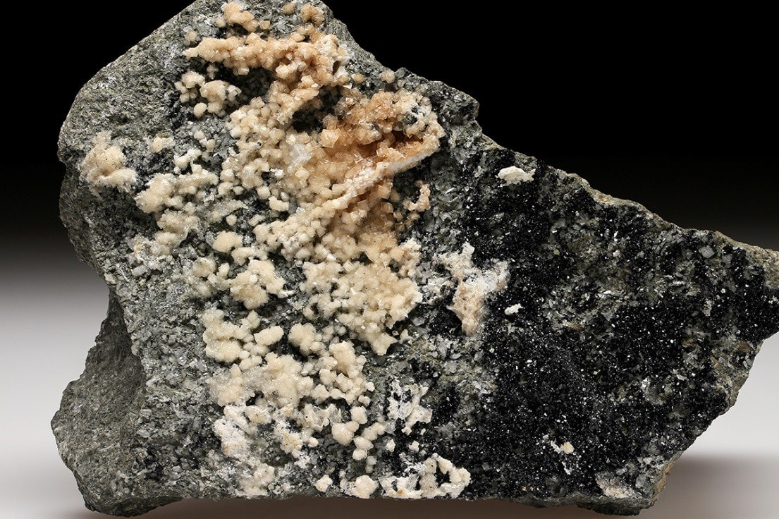
Tan calcite and black babintonite crystals, with minor, broken white apophyllite-(K) between the two just right of center. Specimen 13 x 10 cm. H. Moritz photo.

Specimen 1661
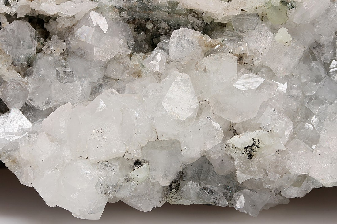
Colorless to white apophyllite-(K) crystals. FOV 7 cm. H. Moritz photo.
Specimen 1873
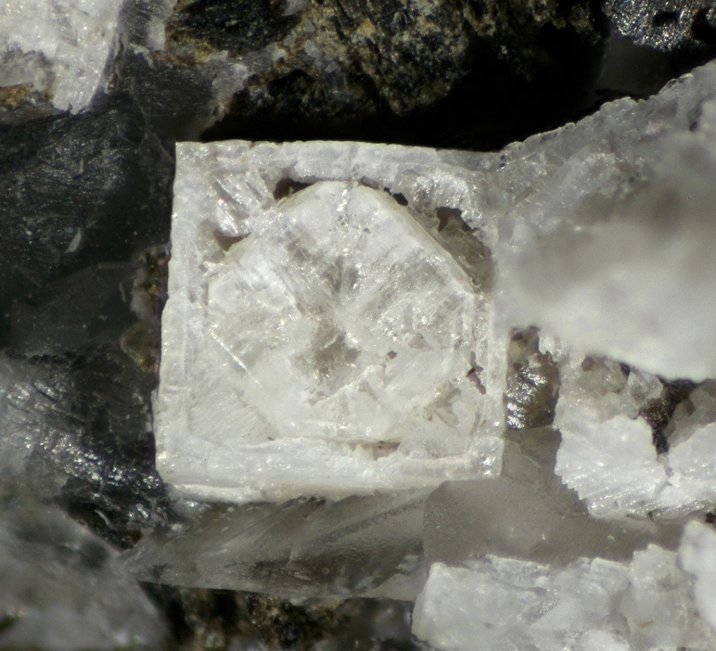
Strange apophyllite-(K) crystal. Field of view: 2.94 mm. H. Moritz photo.
Specimen 3696
Acicular mesolite with small white apophyllite-(K) crystals. Field of view: 6.5 cm. H. Moritz photo.